728x90

понедельник, 1 ноября 2010 г.

Twisted sheets.

X-ray diffraction studies have shown that β pleated sheets are usually not flat but are twisted. In a twisted sheet the individual polypeptide chains make a shallow left-handed helix. However, when successive carbonyl groups are viewed along the direction of the chain, a right-handed twist is seen. Such twisted β sheets are often found in the globular proteins. An example is the “nucleotide-binding” domain of a dehydrogenase enzyme. The twist of the sheet is seen clearly in this stereoscopic view. When such chains are associated into β sheets, whether parallel or antiparallel, and are viewed in a direction perpendicular to the chains and looking down the edge of the sheet, a left-handed “propeller” is seen. Such a propeller is visible in the drawing of carboxypeptidase.
The cause of the twist in β sheets appears to lie in noncovalent interactions between hydrogen atoms on the β-carbon atoms of side chains and the peptide backbone atoms. For side chains of most L- amino acids these interactions provide a small tendency towards the observed right-handed twist. Nonplanarity in the amide groups may also contribute. Interstrand interactions seem to be important.

A “ribbon” drawing of the 307- residue proteinhydrolyzing enzyme carboxypeptidase A. In this type of drawing wide ribbons are used to show β strands and helical turns while narrower ribbons are used for bends and loops of the peptide chains. The direction from the N terminus to C terminus is indicated by the arrowheads on the β strands. No individual atoms are shown and side chains are omitted. Courtesy of Jane Richardson.

четверг, 28 октября 2010 г.

Pleated sheets.

While a fully extended polyglycine chain is possible, the side chains of other amino acids cannot be accommodated without some distortion of the structure. Thus, the peptide chains in silk fibroin have a repeat distance of 0.70 nm compared with the 0.72 nm for the fully extended chain. Pauling and Corey108 showed that this shortening of the chain could result from rotation of angle φ by ∼40° (to –140°) and rotation of ψ in the opposite direction by ∼45° (to +135°) to give a slightly puckered chain. The resulting multichain structure is known as a pleated sheet. As in this figure, both parallel and antiparallel strands are often present in a single β sheet within a protein.
Straight (left) and twisted (right) peptide chains in extended β conformations. From Chothia.


(A) Stereoscopic view of the nucleotide binding domain of glyceraldehyde phosphate dehydrogenase. The enzyme is from Bacillus stearothermophilus but is homologous to the enzyme from animal sources. Residues are numbered 0–148. In this wire model all of the main chain C, O, and N atoms are shown but side chains have been omitted. The large central twisted β sheet, with strands roughly perpendicular to the page, is seen clearly; hydrogen bonds are indicated by dashed lines. Helices are visible on both sides of the sheet. The coenzyme NAD+ is bound at the end of the β sheet toward the viewer. Note that the two phosphate groups in the center of the NAD+ are H-bonded to the N terminus of the helix beginning with R10. From Skarzynski et al. (B) Structural formula for NAD+.

воскресенье, 24 октября 2010 г.

The Extended Chain Structures

As was first pointed out by Pauling and Corey, an important structural principle is that within proteins the maximum possible number of hydrogen bonds involving the C=O and N–H groups of the peptide chain should be formed. One simple way to do this is to line up fully extended chains (φ = ψ = 180°) and to form hydrogen bonds between them. Such a structure exists for polyglycine and resembles that. Notice that on the left side of this figure, the adjacent chains run in opposite directions; hence, the term antiparallel structure. The antiparallel arrangement not only gives the best hydrogen bond formation between chains but also permits a single chain to fold back on itself giving a compact hairpin loop.

The extended chain β pleated sheet structures. (A) Stereoscopic drawing without atomic symbols. (B) Drawing with atomic symbols. At the left is the antiparallel structure. The 0.70 nm spacing is slightly decreased from the fully extended length. The amino acid side chains (R) extend alternately above and below the plane of the “accordion pleated” sheet. The pairs of linear hydrogen bonds between the chains impart great strength to the structure. The chain can fold back on itself using a “β turn” perpendicular to the plane of the pleated sheet. The parallel chain structure (right side) is similar but with a less favorable hydrogen bonding arrangement. Arrows indicate chain directions.

среда, 20 октября 2010 г.

Conformations of Polypeptide Chains

To understand how a polypeptide chain folds we need to look carefully at the possible conformations of the peptide units. Since each peptide unit is nearly planar, we can think of a polypeptide as a chain of flat units fastened together. Every peptide
unit is connected to the next by the α-carbon of an amino acid. This carbon provides two single bonds to the chain and rotation can occur about both of them (except in the cyclic amino acid proline). To specify the conformation of an amino acid unit in a polypeptide chain, we must describe the torsion angles about both of these single bonds. These angles are indicated by the symbols (phi) and (psi) and are assigned the value 180° for the fully extended chain. Each angle is taken as zero for the impossible conformation in which the two chain ends are in the eclipsed conformation. By the same token, the torsion angle (omega) around the C–N bond of the amide is 0° for a planar cis peptide linkage and 180° for the usual trans linkage.
Since both φ and ψ can vary for each residue in a protein, there are a large number of possible conformations. However, many are excluded because they bring certain atoms into collision. This fact can be established readily by study of molecular models.


Some idealized shapes that a 34.5 kDa protein molecule of 300 amino acids might assume.

Two peptide units in the completely extended β conformation. The torsion angles φi, ψi, and ωi are defined as 0° when the main chain atoms assume the cis or eclipsed conformation. The angles in the completely extended chain are all 180°. The distance from one α carbon atom (Cα) to the next in a peptide chain is always 0.38 nm, no matter how the chain is folded.


Using a computer, it is possible to study the whole range of possible combinations of φ and ψ. This has been done for the peptide linkage by Ramachandran. The results are often presented as plots of φ vs ψ (Ramachandran plots or conformational maps)
in which possible combinations of the two angles are indicated by blocked out areas. The original Ramachandran plots were made by representing the atoms as hard spheres of appropriate van der Waals radii. This map was calculated for poly-L-alanine but it would be very similar for most amino acids.
The upper area contains the pairs of torsion angles for the extended structures as well as for collagen. The lower area contains allowed conformations for the right-handed
helices. Most of the observed conformations of peptide units in a real protein fall into these regions. Glycyl residues are an exception. Since glycine has no β-carbon atom, the conformations are less restricted. Out of nearly 1900 non-glycine residues in well-determined protein structures, 66 were found in disallowed areas of the Ramachandran diagram. These were often accommodated by local distortions in bond angles. The positions at which such steric strain occurs are often in regions concerned with function.104b One residue, which lies in a disallowed region. It is located adjacent to the coenzyme site. The possible conformations of proline residues are limited. The angle φ is always –60 ± 20°, while ψ for the residue adjacent to the proline N can be either ~150° or ~ –30°. Typical φ, ψ angles for some regular eptide structures are given.
Potential energy distribution in the φ–ψ plane for a pair of peptide units with alanyl residues calculated using potential parameters of Scheraga and Flory. Contours are drawn at intervals of 1 kcal (4.184 kJ) per mol going down from 0 kcal per mol. The zero contour is dashed. From Ramachandran et al. The points marked x are for the four ideal structures: twisted β structure (β), collagen (C), right-handed α helix (αR), and the less favored left-handed α helix (αL).

суббота, 16 октября 2010 г.

The Architecture of Folded Proteins

All proteins are made in the same way but as the growing peptide chains peel off from the ribosome, each of the thousands of different proteins in a living cell folds into its own special tertiary structure. The number of possible conformations of a protein chain is enormous. Consider a 300-residue polypeptide which could stretch in fully extended form for ∼100 nm. If the chain were folded back on itself about 13 times it could form a 7-nm square sheet about 0.5 nm thick. The same polypeptide could form a thin helical rod 45 nm long and ∼1.1 nm thick. If it had the right amino acid sequence it could be joined by two other similar chains to form a collagen-type triple helix of 87 nm length and about 1.5 nm diameter. The highly folded globular proteins vary considerably
in the tightness of packing and the amount of internal water of hydration. However, a density of ∼1.4 g cm–3 is typical. With an average mass per residue of 115 Da our 300-residue polypeptide would have a mass of 34.5 kDa or 5.74 x 10⁻²⁰ g and a volume of 41 nm3. This might be approximated by a cube 3.45 nm in width, a “brick” of dimensions 1.8 x 3.6 x 6.3 nm, or a sphere of diameter 4.3 nm. Although protein molecules are usually very irregular in shape, for purposes of calculation idealized ellipsoid and rod shapes are often assumed.
It is informative to compare these dimensions with those of the smallest structures visible in cells; for example, a bacterial flagellum is ∼13 nm in diameter and a cell membrane ∼8–10 nm in thickness. Bricks of the size of the 300-residue polypeptide could be used to assemble a bacterial flagellum or a eukaryotic microtubule. Helical polypeptides may extend through cell membranes and project on both sides, while a globular protein of the same chain length may be almost completely embedded in the membrane.

вторник, 12 октября 2010 г.

PROTEINS OF BLOOD PLASMA

Among the most studied of all proteins are those present in blood plasma. Their ready availability and the clinical significance of their study led to the early development of electrophoretic separations. Electrophoresis at a pH of 8.6 (in barbital buffer) indicated six main components. The major and one of the fastest moving proteins is serum albumin. Trailing behind it are the 1-, 2-, and -globulins, fibrinogen, and -globulins. Each of these bands consists of several proteins and two-dimensional separation by electrophoresis and isoelectric focusing reveals over 30 different proteins.e Many of these contain varying numbers of attached carbohydrate units and appear as families of spots.
Fractionation of large quantities of plasma together with immunochemical assays has led to identification of over 200 different proteins. Sixty or more are enzymes, some in very small quantitites which may have leaked from body cells. Normally plasma contains 5.7 – 8.0 g of total protein per 100 ml (~1 mM). Albumin accounts for 3.5 – 4.5 g/100 ml. An individual’s liver synthesizes about 12 g each day. Next most abundant are the immunoglobulins. One of these (IgG or γ-globulin) is present to the extent of 1.2–1.8 g/100 ml. Also present in amounts greater than 200 mg per 100 ml are - and -lipoproteins, the 1 antitrypsin, 2-macroglobulin, haptoglobin, transferrin, and fibrinogen.
Plasma proteins have many functions. One of them, fullfilled principally by serum albumin, is to impart enough osmotic pressure to plasma to match that of the cytoplasm of cells. The heart-shaped human serum albumin consists of a single 65 kDa chain of 585 amino acid residues coiled into 28 helices. Three homologous repeat units or domains each contain six disulfide bridges, suggesting that gene duplication occurred twice during the evolution of serum albumins. The relatively low molecular mass and high density of negative charges on the surface make serum albumin well adapted for the role of maintaining osmotic pressure. However, serum albumin is not essential to life.


Over 50 mutant forms have been found and at least 30 persons have been found with no serum albumin in their blood. These analbuminemic individuals are healthy and have increased concentrations of other plasma proteins.
A second major function of plasma proteins is transport. Serum albumin binds to and carries many sparingly soluble metabolic products, including fatty acids, tryptophan, cysteine, steroids, thyroid hormones, Ca2+, Cu2+, Zn2+, other metal ions, bilirubin, and various drugs. There are also many more specialized transporter proteins. Transferrin carries iron and ceruloplasmin (an α2 globulin) transports copper. Transcortin carries corticosteroids and progesterone, while another protein carries sex hormones. Retinol-binding protein carries vitamin A and cobalamin-binding proteins vitamin B12. Hemopexin carries heme to the liver, where the iron can be recovered.j Haptoglobin binds hemoglobin released from broken red cells and also assists in the recycling of the iron in the heme.k Lipoproteins carry phospholipids, neutral lipids, and cholesterol esters. Most of the mass of these substances is lipid.
Immunoglobulins, α1-trypsin inhibitor and 2-macroglobulin, ten or more blood clotting factors; and proteins of the complement system all have protective functions that are discussed elsewhere in this book. Hormones, many of them proteins, are present in the blood as they are carried to their target tissues. Many serum proteins have unknown or poorly understood functions. Among these are the acute phase proteins, whose concentrations rise in response to inflammation or other injury.

пятница, 8 октября 2010 г.

Polypeptides

The chain formed by polymerization of amino acid molecules provides the primary structure of a protein. Together with any covalent crosslinkages and other modifications, this may also be called the covalent structure of the protein. Each monomer unit in the chain is known as an amino acid residue. This term acknowledges the fact that each amino acid has lost one molecule of H2O during polymerization. To be more precise, the number of water molecules lost is one less than the number of residues. Peptides are named according to the amino acid residues present and beginning with the one bearing the terminal amino group. Thus, L-alanyl-L-valyl-L-methionine has the following structure:


Like amino acids, this tripeptide is a dipolar ion. The same structure can be abbreviated Ala-Val-Met or, using one-letter abbreviations, AVM. It is customary in describing amino acid sequences to place the aminoterminal (N-terminal) residue at the left end and the carboxyl-terminal (C-terminal) residue at the right end. Residues are numbered sequentially with the N-terminal residue as 1.
The sequence of amino acid units in a protein is always specified by a gene. The sequence determines how the polypeptide chain folds and how the folded protein functions. For this reason much effort has gone into “sequencing,” the determination of the precise order of amino acid residues in a protein. Sequences of several hundreds of thousands of proteins and smaller peptides have been established and the number doubles each year.

(A) The complete amino acid sequence of the cytoplasmic enzyme aspartate aminotransferase from pig heart. The peptide has the composition Lys19, His8, Arg26, (Asp + Asn)42, Ser26, Thr26, (Glu + Gln)41, Pro24, Gly28, Ala32, Cys5, Val29, Met6, Ile19, Leu38, Tyr12, Phe23, Trp9. The molecular mass is 46.344 kDa and the complete enzyme is a 93.147-kDa dimer containing two molecules of the bound coenzyme pyridoxal phosphate attached to lysine-258 (enclosed in box).85,86 (B) A stereoscopic view of a complete enzyme molecule which contains two identical subunits with the foregoing sequence. Coordinates from Arthur Arnone. In this “wire model” all the positions of all of the nearly 7000 atoms that are heavier than hydrogen are shown. The > 8000 hydrogen atoms have been omitted. The view is into the active site of the subunit on the right. The pyridoxal phosphate and the lysine residue to which it is attached are shown with heavy lines. The active site of the subunit to the left opens to the back side as viewed here. The drawing may be observed best with a magnifying viewer available from Abrams Instrument Corp., Lansing, Michigan or Luminos Photo Corp., Yonkers, New York. However, with a little practice, it is possible to obtain a stereoscopic view unaided. Hold the book with good illumination about 20–30 cm from your eyes. Allow your eyes to relax as if viewing a distant object. Of the four images that are visible, the two in the center can be fused to form the stereoscopic picture. Drawings by program MolScript (Kraulis, 1991).


Most of these have been deduced from the sequences of nucleotides in DNA. Sequences of some small peptide hormones and antibiotics. The molecular mass of a protein can be estimated from the chain length by assuming that each residue adds 100–115 Da.
The amino acid composition varies greatly among proteins. A typical globular protein contains all or most of the 20 amino acids. The majority are often present in roughly similar amounts but His, Cys, Met, Tyr, and Trp tend to be less abundant than the others. Specialized proteins sometimes have unusual amino acid compositions. For example, collagen of connective tissue contains 33 mole% glycine and 21% of proline + hydroxyproline residues; the major proteins of saliva contain 22% of glutamate + glutamine and 20–45% proline. Cell walls of plants contain both high proline and high glycine polypeptides. One from petunias is 67% glycine. Silk fibroin contains 45% glycine and 29% alanine. A DNA repair protein of yeast has 13 consecutive aspartate residues. The tough eggshell (chorion) of the domesticated silkmoth Bombyx mori contains proteins with ∼30% cysteine. Many proteins consist, in part, of repeated short sequences. For example, the malaria-causing Plasmodium falciparum in its sporozoite stage is coated with a protein that contains 37 repeats of the sequence NANP interspersed with 4 repeats of NDVP. These two sequences have been indicated with single-letter abbreviations for the amino acids.
With a large number of protein and DNA sequences available, it has become worthwhile to compare sequences of the same protein in different species or of different proteins within the same or different species. Computer programs make it possible to recognize similarities and homologies between sequences even when deletions and insertions have occurred. The term homology has the precise biological definition “having a common evolutionary origin,” but it is often used to describe any close similarity in sequence. Among a pair of homologous proteins, a change at a given point in a sequence may be either conservative, meaning that a residue of similar character (large, small, positively charged, nonpolar, etc.) has been substituted, or it may be nonconservative.

понедельник, 4 октября 2010 г.

The Peptide Unit

The very ability of a protein to exist as a complex three-dimensional structure depends upon the properties of the amide linkages between the amino acid units. Many of these properties follow from the fact that an amide can be viewed as a resonance hybrid of the following structures. Because of the partial doublebond character, the C–N bond is shorter than that of a normal single bond and the C=O bond is lengthened.


The observed lengths in nanometers determined by X-ray diffraction measurements are given. The partial double-bond character of the C–N bond has important consequences. The peptide unit is nearly planar as is indicated by the dashed parallelogram.
However, the bonds around the nitrogen retain some pyramidal. Even more important is the fact that there is flexibility. As a result, the torsion angle ω may vary over a range of ± 15° or even more from that in the planar state. The resonance stabilization of the amide linkage is thought to be about 85 kJ/mol. Rotation around the C–N bond through 90° would be expected to require about this much energy. This fact immediately suggests a way in which proteins may sometimes be able to store energy—by having one or more peptide units twisted out of complete planarity.
Dimensions of the peptide linkage. Interatomic distances in nm, including the hydrogen bond length to an adjacent peptide linkage, are indicated. The atoms enclosed by the dotted lines all lie approximately in a plane. However, as indicated in the lower drawing, the nitrogen atom tends to retain some pyramidal character.


An important effect of the resonance of the amide linkage is that the oxygen atom acquires some negative charge and the NH group some positive charge. Some of the positive charge is usually depicted as residing on the nitrogen, but some is found on the hydrogen atom. The latter can be pictured as arising from a contribution of a fourth resonance form that contains no bond to hydrogen.


Nevertheless, this picture is inadequate. Various evidence indicates that the nitrogen actually carries a net negative charge.
The positive and negative ends of the dipoles in the amide group tend to associate to form strong hydrogen bonds. These hydrogen bonds together with the connecting amide linkages can form chains that may run for considerable distances through proteins. The tendency for cooperativity in hydrogen bond formation may impart unusual stability to these chains. As with individual amide linkages, these chains of hydrogen-bonded amides can also be thought of as resonance hybrids:


The two structures pictured are extreme forms, the true structure being something in between. In the lower form, rotation about the C–N bond would be permitted but then the charge separation present in the upper structure would no longer exist. Thus, the hydrogen bonds would be weakened. We can conclude that if an amide linkage in such a chain becomes twisted, the hydrogen bonds that it forms will be weakened. If there is cooperativity, the hydrogen bonds will all be strongest when there is good planarity in all of the amides in the chain.
Amides have very weak basic properties and protonation is possible either on the oxygen (A) or on the nitrogen (B).


The pKa values for such protonation are usually less than zero, but it is possible that a correctly placed acidic group in a protein could protonate either oxygen or nitrogen transiently during the action of a protein. Protonation on oxygen would strengthen hydrogen bonds from the nitrogen whereas protonation on nitrogen would weaken hydrogen bonds to oxygen and might permit rotation. The amide group has a permanent dipole moment of 3.63 debyes oriented as follows:


Here the arrow points toward the positive end of the dipole.

четверг, 30 сентября 2010 г.

Acidic and Basic Side Chains

The side chains of aspartic and glutamic acids carry negatively charged carboxylate groups at pH 7 while those of lysine and arginine carry the positively charged –NH3 + and guanidinium ions, respectively.

At pH 7 the weakly basic imidazole group of histidine may be partially protonated. Both the –SH group of cysteine and the phenolic –OH of tyrosine are weakly acidic and will dissociate and thereby acquire negative charges at a sufficiently high pH.
The number of positive and negative charges on a protein at any pH can be estimated approximately from the acid dissociation constants (usually given as pKa values) for the amino acid side chains. However, pKa values of buried groups are often greatly shifted from these, especially if they associate as ion pairs. In addition, many proteins have free amino and carboxyl-terminal groups at the opposite ends of the peptide chain. These also participate in acid–base reactions with approximately the following pKa values.

terminal --, pKa = 3.6–3.7
terminal --NH3+, pKa = 7.5–7.9

The acid–base properties of an amino acid or of a protein are described by titration curves of the type. In these curves the number of equivalents of acid or base that have reacted with an amino acid or protein that was initially at neutral pH are plotted against pH. The net negative or positive electrical charge on the molecule can be read directly from the curves. Both the net electrical charges and the distribution of positively and negatively charged groups are often of crucial importance to the functioning of a protein.

воскресенье, 26 сентября 2010 г.

Properties of -Amino Acids

The amino acids have in common a dipolar ionic structure and a chiral center. They are differentiated, one from another, by the structures of their side chain groups, designated R in the foregoing formulas. These groups are of varying size and chemical structure. The side chain groups fill much of the space in the interior of a protein molecule and also protrude from the external surfaces of the protein where they determine many of the chemical and physical properties of the molecule.
Show the structures of the side chains of the amino acids commonly found in proteins. The
complete structure is given for proline. Both the threeletter abbreviations and one-letter abbreviations used in describing sequences of amino acids in proteins are also given in this table. Amino acids of groups a–c plus phenylalanine and methionine are sometimes grouped together as nonpolar. They tend to be found in a hydrophobic environment on the inside of a protein molecule. Groups f and i contain polar, charged side chains which usually protrude into the water surrounding the protein. The rest are classified as polar but noncharged.
To get acquainted with amino acid structures, learn first those of glycine, alanine, serine, aspartic acid, and glutamic acid. The structures of many other amino acids can be related to that of alanine (R=CH3) by replacement of a β hydrogen by another group. Metabolic interrelationships will make it easier to learn structures of the rest of the amino acids later.

Since the –COOH groups of glutamic and aspartic acids are completely dissociated to –COO– at neutral pH, it is customary in the biochemical literature to refer to these amino acids as glutamate and aspartate without reference to the nature of the cation or cations present as counter ions. Such “-ate” endings are also used for most other acids (e.g., malate, oxaloacetate, phosphate, and adenylate) and in names of enzymes (e.g., lactate
dehydrogenase).
During the formation of polypeptides, the α-amino and carboxyl groups of the amino acids are converted into the relatively unreactive and uncharged amide (peptide)
groups except at the two chain termini. In many cases the terminal amino and carboxyl groups are also converted within cells into uncharged groups (Chapter 10). Immediately
after the protein is synthesized its terminal carboxyl group is often converted into an amide. The N terminus may be acetylated or cyclized to a pyroglutamyl group. Sometimes a cyclic peptide is formed.
The properties of polypeptides and proteins are determined to a large extent by the chemistry of the side chain groups, which may be summarized briefly as follows. Glycine in a peptide permits a maximum of conformational mobility. The nine relatively nonpolar amino acids–alanine, valine, leucine, isoleucine, proline, methionine, phenylalanine, tyrosine, and tryptophan– serve as building blocks of characteristic shape. Tyrosine and tryptophan also participate in hydrogen bonding and in aromatic: aromatic interactions within proteins.
Much of the chemistry of proteins involves the side chain functional groups –OH, –SH, –COO–, –NH3 +, and imidazole and the guanidinium group of arginine. The side chains of asparagine and glutamine both contain the amide group CONH2, which is relatively inert chemically but which can undergo hydrogen-bonding interactions. The amide linkages of the polypeptide backbone must also be regarded as important functional groups. Most polar groups are found on the outside surfaces of proteins where they can react chemically in various ways. When inside proteins they form H-bonds to the peptide backbone and to other polar groups.

среда, 22 сентября 2010 г.

Amino Acids and Peptides

Twenty α-amino acids are the monomers from which proteins are made. These amino acids share with other biochemical monomers a property essential to their role in polymer formation: They contain at least two different chemical groups able to react with each other to form a covalent linkage. In the amino acids these are the protonated amino (NH3 +) and carboxylate (COO–) groups. The characteristic linkage in the protein polymer is the peptide (amide) linkage whose formation can be imagined to occur by the splitting out of water between the carboxyl of one amino acid and the amino group of another.
This equation is not intended to imply a mechanism for peptide synthesis. The equilibrium position for this reaction in an aqueous solution favors the free amino acids rather than the peptide. Therefore, both biological and laboratory syntheses of peptides usually do not involve a simple splitting out of water. Since the dipeptide still contains reactive carboxyl and amino groups, other amino acid units can be joined by additional peptide linkages to form polypeptides. These range from short-chain oligomers to polymers of from ∼50 to several thousand amino acid units, the proteins.

суббота, 18 сентября 2010 г.

Hydrophobic Interactions

Fats, hydrocarbons, and other materials whose molecules consist largely of nonpolar groups have a low solubility in water and a high solubility in nonpolar solvents. Similarly, the long alkyl groups of fatty acid esters aggregate within membranes and nonpolar side chains of proteins are often packed together in the centers of protein molecules. Because it is as if the nonpolar groups “fear” water, this is known as the hydrophobic effect. The terms hydrophobic forces, hydrophobic interactions, and hydrophobic bonding have also been used. However, the latter term can be misleading because the hydrophobic effect arises not out of any special attraction between nonpolar groups but primarily from the strong internal cohesion of the hydrogen-bonded water structure.
–CH2–(water) --→–CH2–(hexane)
ΔG° = –3.8 kJ/mol


This equation is a quantitative statement of the fact that the CH2 group prefers to be in a nonpolar environment than to be surrounded by water. A similar Gibbs energy change would be expected to accompany the bringing together of a methylene unit from a small molecule and a hydrophobic surface on a protein molecule. However, in the latter case the accompanying What causes the decrease in Gibbs energy when nonpolar groups associate in water? Jencks60 suggested that we think of the transfer of a nonpolar molecule from a nonpolar solvent into water in two steps: (1) Create a cavity in the water of about the right size to accommodate the molecule. Since many hydrogen bonds will be broken, the Gibbs energy of cavity formation will be high. It will be principally an enthalpy (ΔH) effect. (2) Allow the water molecules in the solvent to make changes in their orientations to accommodate the nonpolar molecule that has been placed in the cavity. The water molecules can move to give good van der Waals contacts and also reorient themselves to give the maximum number of hydrogen bonds. Since hydrogen bonds can be formed in many different ways in water, there may be as many or even more hydrogen bonds after the reorientation than before. This will be true especially at low temperature where most water exists as large icelike clusters. For dissolved hydrocarbons, the enthalpy of formation of the new hydrogen bonds often almost exactly balances the enthalpy of creation of the cavity initially so that ΔH for the overall process (transfer from inert solvent into water) is small. For the opposite transfer ΔH° is usually a small positive number for aliphatic hydrocarbons and is nearly zero for aromatic hydrocarbons.
Since ΔG° = ΔH°– TΔS°, it follows that the negative value of ΔG° for hydrophobic interactions must result from a positive entropy change, which may arise from the restricted mobility of water molecules that surround dissolved hydrophobic groups. When two hydrophobic groups come together to form a “hydrophobic bond,” water molecules are freed from the structured region around the hydrophobic surfaces and the entropy increases. The ΔS°. Attempts have been made to relate this value directly to the increased number of orientations possible for a water molecule when it is freed from the structured region. However, interpretation of the hydrophobic effect is complex and controversial.
The formation constant Kf for hydrophobic associations often increases with increasing temperature. This is in contrast to the behavior of Kf for many association reactions that involve polar molecules and for which ΔH° is often strongly negative (heat is released). An example of the latter is the protonation of ammonia in an aqueous solution

NH3 + H+ → NH4+
ΔH° = –52.5 kJ/mol


Since R1nKf = –ΔG°/T = –ΔH°/T + ΔS°, Kf decreases with increasing temperature if ΔH° is negative. Because for a hydrophobic interaction with a positive value of ΔH° Kf increases with increasing temperature, an increase in stability at higher temperatures is sometimes used as a criterion for hydrophobic bonding. However, this criterion does not always hold. For example, base stacking interactions in polynucleotides, whose strength does not increase with increasing temperature, are still thought to be hydrophobic.
The water molecules that are in immediate contact with dissolved nonpolar groups are partially oriented. They form a cagelike structure around each hydrophobic group. When particles surrounded by such hydration layers are 1–2 nm apart, they sometimes experience either a fairly strong repulsion or an enhanced attraction caused by these hydration layers Direct experimental measurements have shown that these effects extend to distances of 10 nm and can account for the previously mentioned long-range van der Waals forces.
Various efforts have been made to develop scales of hydrophobicity that can be used to predict the probability of finding a given amino acid side chain buried within a protein or in a surface facing water. A new approach has been provided by the study of mutant proteins. For example, deletion of a single –CH2– group from an interior hydrophobic region of a protein was observed to decrease the stability of the protein by 4.6 kJ/mol.

пятница, 17 сентября 2010 г.

Hydration of Polar Molecules and Ions

Water molecules are able to hydrogen bond not only to each other but also to polar groups of dissolved compounds. Thus, every group that is capable of forming a hydrogen bond to another organic group is also able to form hydrogen bonds of a somewhat similar strength with water. For this reason, hydrogen bonding is usually not a significant force in holding small molecules together in aqueous solutions. Polar molecules that stick together through hydrogen bonding when dissolved in a nonpolar solvent often do not associate in water. How then can biochemists assert that hydrogen bonding is so important in biochemistry? Part of the answer is that proteins and nucleic acids can be either properly folded with hydrogen bonds
formed internally or denatured with hydrogen bonds from those same groups to water. The Gibbs energy change between these two states is small.
Every ion in an aqueous solution is surrounded by a shell of oriented water molecules held by the attraction of the water dipoles to the charged ion. The hydration of ions has a strong influence on all aspects of electrostatic interactions and plays a dominant role in determining such matters as the strength of acids and bases, the Gibbs energy of hydrolysis of ATP, and the strength of bonding of metal ions to negatively charged groups. For example, the previously considered interaction between carboxylate and calcium ions would be much weaker if both ions retained their hydration shells.
Consider the following example. ΔG° for dissociation of acetic acid in water is +27.2 kJ/mol. The enthalpy change ΔH° for this process is almost zero (–0.1 kJ/mol) and ΔS° is consequently –91.6 J K–1. This large entropy decrease reflects the increased amount of water that is immobilized in the hydration spheres of the H+ and acetate– ions formed in the dissociation reaction. In contrast, dissociation of NH4 + to NH3 and H+ converts one positive ion to another. ΔH° is large (+52.5 kJ/mol) but the entropy change ΔS° is small.
Although effects of hydration are important in almost all biochemical equilibria, they are difficult to assess quantitatively. It is hard to know how many molecules of water are freed or immobilized in a given reaction. Charged groups in proteins are often hydrated. However, if they are buried in the interior of the protein, they may be solvated by polarizable protein side chain groups such as –OH or by backbone or side chain amide groups.

среда, 8 сентября 2010 г.

The Structure and Properties of Water

Water is the major constituent of cells and a remarkable solvent whose chemical and physical properties affect almost every aspect of life. Many of these properties are a direct reflection of the fact that most water molecules are in contact with their neighbors entirely through hydrogen bonds.Water is the only known substance for which this is true.
In ordinary ice all of the water molecules are connected by hydrogen bonds, six molecules forming a hexagonal ring resembling that of cyclohexane. The structure is extended in all directions by the formation of additional hydrogen bonds to adjacent molecules. As can be seen in this drawing, the molecules in ice assume various orientations in the hexagonal array, and frequently rotate to form their hydrogen bonds in different ways. This randomness remains as the temperature is lowered, and ice is one of few substances with a residual entropy at absolute zero. Ice is unusual also in that the molecules do not assume closest packing in the crystal but form an open structure. The hole through the middle of the hexagon and on through the hexagons lying below it is ∼0.06 nm in diameter.
The short hydrogen-bond length (averaging 0.276 nm) in ice indicates of strong bonding. The heat of sublimation (ΔH°) of ice is –48.6 kJ/mol. If the van der Waals dispersion energy of –15 kJ/mol is subtracted from this, the difference of –34 kJ/mol can be attributed entirely to the hydrogen bonds—two for each molecule. Their average energy is 17 kJ/mol apiece. However, some of the hydrogen bonds are stronger and others weaker than the average.

Six water molecules in the lattice of an ice crystal. The hydrogen bonds, which connect protons with electron pairs of adjacent molecules, are shown as dashed lines.


In a gaseous water dimer the hydrogen bond is linear, a fact that suggests some covalent character.

Its length is distinctly greater than that in ice. This is one of a number of pieces of evidence suggesting cooperativity in formation of chains of hydrogen bonds. Consider the following three trimers for which theoretical calculations have predicted the indicated hydrogen bond energies. In the first case the central water molecule donates two protons for hydrogen-bond formation; in the second it accepts the protons. In the third case it is both an electron acceptor and a donor. The OH dipoles are oriented “head to tail” and the hydrogen bonds are stronger than in the other


cases. Long chains of similarly oriented hydrogen bonds exist in ice and this may account for the short hydrogen bond lengths. Closed rings of hydrogen bonds oriented to give a maximum cooperative effect also exist in liquid water clusters and within proteins, carbohydrates, and nucleic acids.
The nature of liquid water is still incompletely understood,but we know that water contains icelike clusters of molecules that are continually breaking up and reforming in what has been called a “flickering cluster” structure. Judging by the infrared spectrum of water, about 10% of the hydrogen bonds are broken when ice melts.41 A similar conclusion can be drawn from the fact that the heat of melting of ice is –5.9 kJ/mol. It has been estimated that at 0°C the average cluster contains about 500 water molecules.41 At 50°C there are over 100 and at the boiling point about 40. Although most molecules in liquid water are present in these clusters, the hydrogen bonds are rapidly broken and reformed in new ways, with the average lifetime of a given hydrogen bond being ∼10(–12) s.

воскресенье, 5 сентября 2010 г.

Hydrogen Bonds

One of the most important weak interactions between biologically important molecules is the hydrogen bond (H-bond). These “bonds” are the result of electrostatic attraction caused by the uneven distribution of electrons within covalent bonds. For example, the bonding electron pairs of the H–O bonds of water molecules are attracted more tightly to the oxygen atoms than to the hydrogen atoms. A small net positive charge is left on the hydrogen and a small net negative charge on the oxygen. Such polarization of the water molecules can be indicated in the following way:


Here the δ+ and δ– indicate a fraction of a full charge present on the hydrogen atoms and on the nonbonded electron pairs of the oxygen atom, respectively. Molecules such as H2O, with strongly polarized bonds, are referred to as polar molecules and functional groups with such bonds as polar groups. They are to be contrasted with such nonpolar groups as the –CH3 group in which the electrons in the bonds are nearly equally shared by carbon and hydrogen.
A hydrogen bond is formed when the positively charged end of one of the dipoles (polarized bonds) is attracted to the negative end of another dipole. Water molecules tend to hydrogen bond strongly one to another; each oxygen atom can be hydrogen-bonded to two other molecules and each hydrogen to another water molecule. Thus, every water molecule can have up to four hydrogen-bonded neighbors.

A water molecule hydrogen bonded to four other water molecules; note the tetrahedral arrangement of bonds around the central oxygen.


Many groups in proteins, carbohydrates, and nucleic acids form hydrogen bonds to one another and to surrounding water molecules. For example, an imidazole group of a protein can bond to an OH group of an amino acid side chain or of water in the following
ways:


Remember that hydrogen bonds are always formed between pairs of groups, with one of them, often C=O or C=N-, containing the negative end of a dipole and the other providing the proton. The proton acceptor group, often OH or NH and occasionally SH, and even CH in certain structures,donates an unshared pair of electrons. Dashed arrows are sometimes drawn from the hydrogen atom to the electron donor atom to indicate the direction of a hydrogen bond. Do not confuse these arrows with the curved arrows that indicate flow of electrons in organic reactions.
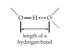
The strength of hydrogen bonds, as measured by the bond energy, varies over the range 10–40 kJ/mol. The stronger the hydrogen bond the shorter its length. Because hydrogen atoms can usually not be seen in X-ray structures of macromolecules, the lengths of
hydrogen bonds are often measured between the surrounding heavy atoms:


A typical —OH- - -O hydrogen bond will have a length of about 0.31 nm; a very strong hydrogen bond may be less than 0.28 nm in length, while weak hydrogen bonds will approach 0.36 nm, which is the sum of the van der Waals contact distances plus the O–H bond length. Beyond this distance a hydrogen bond cannot be distinguished easily from a van der Waals contact.
Hydrogen bonds are strongest when the hydrogen atom and the two heavy atoms to which it is bonded are in a straight line. For this reason hydrogen bonds tend to be linear. However, the dipoles forming the hydrogen bond do not have to be colinear for strong hydrogen bonding: There is some preference for hydrogen bonding to occur in the direction of an unshared electron pair on the oxygen or nitrogen atom.


A linear O–H- - -O hydrogen bond with dipoles at an angle one to another.


Both ammonia, NH3, and the –NH2 groups of proteins are good electron donors for hydrogen bond formation. However, the hydrogen atoms of uncharged –NH2 groups tend to be poor proton donors for H-bonds. Do hydrogen bonds have some covalent character? The answer is controversial.
Hydrogen bonding is important both to the internal structure of biological macromolecules and in interactions between molecules. Hydrogen bonding often provides the specificity necessary to bring surfaces together in a complementary way. Thus, the
location of hydrogen-bond forming groups in surfaces between molecules is important in ensuring an exact alignment of the surfaces.37 The hydrogen bonds do not always have to be strong. For example, Fersht and coworkers, who compared a variety of mutants of an enzyme of known three-dimensional structure, found that deletion of a side chain that formed a good hydrogen bond to the substrate weakened the binding energy by only 2–6 kJ/mol. However, loss of a hydrogen bond to a charged group in the substrate caused a loss of 15–20 kJ/mol of binding energy. Study of mutant proteins created by genetic engineering is now an important tool for experimentally investigating the biological roles of hydrogen bonding.
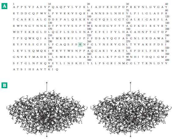
среда, 25 августа 2010 г.

Attraction between Charged Groups (Salt Linkages).

Fixed positive and negative charges attract each other strongly. Consider a carboxylate ion in contact with–NH3 + or with an ion of calcium:

From the van der Waals radii of Table 2-1 and the ionic crystal radius of Ca2+ of 0.10 nm, we can estimate an approximate distance between the centers of positive and negative charge of 0.25 nm in both cases. It is of interest to apply Coulomb’s law to compute the force F between two charged particles which are almost in contact. Let us choose a distance of 0.40 nm.

In this equation r is the distance in meters, q and q’ are the charges in coulombs (one electronic charge = 1.6021 x 10⁻¹⁹ coulombs), ε is the dielectric constant, and F is the force in newtons (N). The force per mole is NF where N is Avogadro’s number.
An uncertainty in this kind of calculation is in the dielectric constant ε, which is 1.0 for a vacuum, about 2 for hydrocarbons, and 78.5 for water at 25°C. If ε is taken as 2, the force for r = 0.40 nm is 4.3 x 10¹⁴ N/mol. The force would be twice as great for the Ca2+ – COO– case. To move two single charges further apart by just 0.01 nm would require 4.3 kJ/mol, a substantial amount of energy. However, if the dielectric constant were that of water, this would be reduced almost 40-fold and the electrostatic force would not be highly significant in binding. It is extremely difficult to assign a dielectric constant for use in the interior of proteins. For charges spaced far apart within proteins the effective dielectric constant is usually as high as 30–60. For closely spaced charges in hydrophobic niches it may be as low as 2–4.
A calculation that is often made is the work required to remove completely two charges from a given distance apart (e.g., 0.40 nm) to an infinite distance.

If ε = 2, this amounts to 174 kJ/mol for single charges at a distance of 0.40 nm; 69 kJ/mol at 1 nm; and only 6.9 kJ/mol at 10 nm, the distance across a cell membrane. We see that very large forces exist between closely spaced charges.
Electrostatic forces are of great significance in interactions between molecules and in the induction of changes in conformations of molecules. For example, attraction between –COO– and –NH3 + groups occurs in interactions between proteins. Calcium ions often interact with carboxylate groups, the doubly charged Ca2+ bridging between two carboxylate or other polar groups. This occurs in carbohydrates such as agarose, converting solutions of these molecules into rigid gels. Individual macromolecules as well as cell surfaces usually carry a net negative charge at neutral pH. This causes the surfaces to repel each other. However, at a certain distance of separation the van der Waals attractive forces will balance the electrostatic repulsion. Protruding hydrophobic groups may then interact and may“tether” bacteria or other particles at a fixed distance, often ~5 nm, from a cell surface.

понедельник, 23 августа 2010 г.

Van der Waals Forces.

All atoms have a weak tendency to stick together,
and because of this even helium liquifies at a low
enough temperature. This is a result of the van der
Waals or “London dispersion forces” which act strongly only at a very short distance. These forces arise from electrostatic attraction between the positively charged nucleus of one atom and the negatively charged electrons of the other. Because nuclei are screened by the electron clouds surrounding them, the force is weak. The energy (enthalpy) of binding of one methylene (–CH2 –) unit into a monomolecular layer of a fatty acid is about –∆H° = 1.7 kJ/mol. 22 Although this is a small quantity, when summed over the 16 or more carbon atoms of a typical fatty acid the binding energy is substantial. When a methylene group is completely surrounded in a crystalline hydrocarbon, its van der Waals energy, as estimated from the heat of sublimation, is 8.4 kJ/mol; that of H2O in liquid water at the melting point of ice is 15 kJ/mol.
While van der Waals forces between individual atoms act over very short distances, they can be felt at surprisingly great distances when exerted by large molecules or molecular aggregates. Forces between very smooth surfaces have been measured experimentally at distances as great as 10 nm and even to 300 nm. However, these “long-range van der Waals forces” probably depend upon layers of oriented water molecules on the plates.

суббота, 21 августа 2010 г.

Forces between Molecules and between Chemical Groups.

The structure of living cells depends very much on the covalent bonds within individual molecules and on covalent crosslinks that sometimes form between molecules. However, weaker forces acting between molecules and between different parts of the same molecule are responsible for many of the most important properties of biochemical substances. These
are described as van der Waals forces, electrostatic forces, hydrogen bonds, and hydrophobic interactions. In the discussion that follows the thermodynamic quantities ∆H, ∆S, and ∆G will be used.

четверг, 19 августа 2010 г.

Tautomerism and Resonance.

Many simple organic compounds exist as mixtures of two or more rapidly interconvertible isomers or tautomeric forms. Tautomers can sometimes be separated one from the other at low temperatures where the rate of interconversion is low. The classic example is the oxo-enol (or keto-enol) equilibrium.

Although usually less stable than the oxo (keto) form, the enol is present in a small amount. It is formed readily from the oxo tautomer by virtue of the fact that hydrogen atoms attached to carbon atoms that are immediately adjacent to carbonyl (C=O) groups are remarkably acidic. Easy dissociation of a proton is a prerequisite for tautomerism. Since most hydrogen atoms bound to carbon atoms do not dissociate readily, tautomerism is unusual unless a carbonyl or other “activating group” is resent.
Since protons bound to oxygen and nitrogen atoms usually do dissociate readily, tautomerism also exists in amides and in ring systems containing O and N.


The tautomerism in is the counterpart of that in the oxo-enol transformation. However, the equilibrium constant for aqueous conditions favors form A very strongly. 2-Pyridone is tautomerized to 2-hydroxypyridine to a greater extent. Pyrimidines and purines can form a variety of tautomers. The existence of form D of is the basis for referring to uracil as dihydroxypyrimidine. However, the di-oxo tautomer A redominates. Pyridoxine (vitamin B6) exists in water largely as the dipolar ionic tautomer B but in methanol as the uncharged tautomer A. In a pair of tautomers, a hydrogen atom always moves from one position to another and the lengths and bond haracter of these bonds also change.
The equilibrium constant for a tautomeric interconversion is simply the ratio of the mole fractions of the two forms; for example, the ratio of enol to oxo forms of acetone12 in water at 25°C is 6.0 x 10⁻⁹, while that for isobutyraldehyde is 1.3 x 10⁻⁴. The ratio of 2-hydroxypyridine to 2-pyridone is about 10–3 in water but increases to 0.6 in a hydrocarbon solvent and to 2.5 in the vapor phase. The ratio of dipolar ion to uncharged pyridoxine is ∼4 at 25°C in water. The ratios of tautomers B, C, and D to the tautomer A of uracil are small, but it is ifficult to measure them quantitatively. These tautomeric ratios are defined for given overall states of rotonation. The constants are independent of pH but will change if the overall state f protonation of the molecule is changed. They may also be altered by changes in temperature or solvent or by binding to a protein or other molecule.
It is important to distinguish tautomerism from resonance, a term used to indicate that the properties of a given molecule cannot be represented by a single valence structure but can be represented as a hybrid of two or more structures in which all the nuclei remain in the same places. Only bonding electrons move to convert one resonance form into another. Examples are the enolate anion, which can be thought of as a hybrid of structures A and B, and the amide linkage, which can be represented by a similar pair of resonance forms.

A double-headed arrow is often used to indicate that two structures drawn are resonance structures rather than tautomers or other separable isomers.
Although they are distinctly different, tautomerism and resonance are related. Thus, the acidity of carbon-bound hydrogens in ketones, which allows formation of enol tautomers, results from the fact that the enolate anion produced by dissociation of one of these hydrogens is stabilized by resonance. Similarly, tautomerism in the imidazole group of the amino acid histidine is related to resonance in the imidazolium cation. Because of this resonance, if a proton approaches structure A of and becomes attached to the lefthand nitrogen atom (Nδ), the positive charge in the resulting intermediate is distributed over both nitrogen atoms. This makes the proton on Nε acidic, permitting it to dissociate to tautomer B.

Since Nσ has sometimes also been called N3, it is best not to use the numerical designations for the nitrogen atoms. The tautomeric ratio of B to A for histidine in
water has been estimated, using 15N- and 13C-NMR, as 5.0 when the α-amino group is protonated and as 2.5 when at high pH it is unprotonated. This tautomerism of the imidazole group is probably important to the function of many enzymes and other proteins; for example, if Nε of structure A is embedded in a protein, a proton approaching from the outside can induce the tautomerism shown with the release of a proton in the interior of the protein, perhaps at the active site of an enzyme. The form protonated on Nδ, which is the minor form in solution, predominates in some positions within proteins.

вторник, 17 августа 2010 г.

Conformations: The Shapes That Molecules Can Assume.

As important to biochemists as configurations, the stable arrangements of bonded atoms, are conformations, the various orientations of groups that are caused by rotation about single bonds. In many molecules such rotation occurs rapidly and freely at ordinary temperatures. We can think of a –CH3 group as a kind of erratic windmill, turning in one direction, then another. However, even the simplest molecules have preferredconformations, and in more complex structures rotation is usually very restricted.
Consider a molecule in which groups A and B are joined by two CH2 (methylene) groups. If A and B are pulled as far apart as possible, the molecule is in its fully extended anti or staggered conformation:


Groups A and B are said to be antiperiplanar (ap) in this conformation. Not only are A and B as far apart as possible but also all of the hydrogen atoms are at their maximum distances one from the other. This can be seen by viewing the molecule down the axis joining the carbon atoms (Newman projection). Rotation of the second carbon atom 180° around the single bond yields the eclipsed conformation in which groups A and B are synperiplanar.


If A and B are large bulky groups they will bump together, attainment of the eclipsed conformation will be almost impossible, and rotation will be severely restricted. Even if A and B are hydrogen atoms (ethane), there will be a rotational barrier in the eclipsed conformation which amounts to ∼12 kJ (3 kcal) per mole because of the crowding of the hydrogen atoms as they pass each other. This can be appreciated readily by examination of space-filling molecular models.
If groups A and B are methyl groups (butane), the steric hindrance between A and B leads to a rotational barrier of ∼25 kJ ( 6 kcal) per mole. The consequence of this simple fact is that in fatty acids and related substances and in polyethylene the chains of CH2 groups tend to assume fully extended zigzag conformations.
In addition to this extended conformation there are two gauche (skewed or synclinal) conformations which are only slightly less stable than the staggered conformation and in which A and B interfere only if they are very bulky. In one of the twogauche conformations B lies to the right of A and in the other to the left of A when viewed down the axis.


These two conformations are related to right-handed and left-handed screws, respectively. The threads on an ordinary right-handed household screw, when viewed down the axis from either end, move backward from left to right in the same fashion as do the groups A and B in the illustration. The angle φ is the torsion angle and is positive for right-handed conformations. Gauche conformations are important in many biological molecules; for example, the sugar alcohol ribitol stacks in crystals in a “sickle” conformation, in which the chain starts out (at the left) in the zigzag arrangement but shifts to a gauche conformation around the fourth carbon atom, thereby minimizing steric interference between the OH groups on the second and fourth carbons.


The complete series of possible conformations is shown
in Fig 1.


Figure 1. Description of conformations about a single bond in the terminology of Klyne and Prelog10,11 using the Newman projection. Group A is on the front atom at the top: the conformation is given for each possible position of group B on the other atom.

In the chain of methylene units, the hydrogen atoms on alternate carbon atoms of the fully extended chain barely touch but larger atoms cannot be accommodated. Thus, when fluorine atoms of van der Waals radius 0.135 nm replace the hydrogen atoms of radius 0.12 nm, a fully extended chain is no longer possible. For this reason the torsion angle in polyfluoroethylene is changed from the 180° of polyethylene to 166°, enough to relieve the congestion but not enough to cause severe eclipsing of the fluorines on djacent carbons. The resulting helical structure is reminiscent of those occurring in proteins and other biopolymers. We see that helix formation can be a natural result of steric hindrance between groups of atoms.

воскресенье, 15 августа 2010 г.

Geometrical isomers.

The RS system also gives an unambiguous designation of geometrical isomers containing a double bond. At each end of the bond, select the group of highest priority. If these two groups lie on the same side of the double bond the configuration is Z (from the German zusammen,“together”); if on opposite sides E (entgegen,“opposite”).


Configurations of amide or ester linkages may also be specified in this manner. This is possible because the C–N bond of an amide has partial double-bond character, as to a lesser extent does the C–O bond to the bridge oxygen in an ester. In this case, assign the lowest priority to the unshared electron pair on the ester bridge oxygen.


An amide of the Z configuration is ordinarily referred to as trans in protein chemistry because the main chain atoms are trans.

пятница, 13 августа 2010 г.

Diastereoisomers.

Whereas compounds with one chiral center exist as an enantiomorphic pair, molecules with two or more chiral centers also exist as diastereoisomers (diastereomers). These are pairs of isomers with an opposite configuration at one or more of the chiral centers, but which are not complete mirror images of each other. An example is L-threonine which has the 2S, 3R configuration. The diastereoisomer with the 2S, 3S configuration is known as L-allo-threonine. L-isoleucine, whose side chain is –CH(CH3) CH2CH3, has the 2S, 3R configuration. It can be called 2(S)- amino-3(R)-methyl-valeric acid but the simpler name L-isoleucine implies the correct configuration at both chiral centers.
Sometimes the subscript s or g is added to a D or L prefix to indicate whether the chirality of a compound is being related to that of serine, the traditional configurational standard for amino acids, or to that of glyceraldehyde. In the latter case the sugar convention (Chapter 4) is followed. In this convention the configurations of the chiral centers furthest from C1 are compared. Ordinary threonine is Ls- or Dg-threonine. The configuration of dextrorotatory (+)-tartaric acid can be described as 2R, 3R, or as Ds, or as Lg.

Biochemical reactions are usually stereospecific and a given enzyme will catalyze reactions of molecules of only a single configuration. A related fact is that proteins ordinarily consist entirely of amino acids of the L series.

среда, 11 августа 2010 г.

The RS notation for configuration.

This notation, devised by Cahn, Ingold, and Prelog, provides an unambiguous way of specifying configuration at any chiral center. It is especially useful for classes of compounds for which no well-established DL system is available. The groups or atoms surrounding the central carbon atom, or other central atom, are ranked according to a prioritysequence. The priority of a group is determined by a number of sequence rules, the first of which is (1) Higher atomic number precedes lower. In the following illustration, the priorities of the groups in D-alanine are indicated by the letters a > b > c > d. The highest priority (a) is assigned to the NH2 groups which contain nitrogen bonded to the central atom. To establish the configuration, the observer views the molecule down the axis connecting the central atom to the group having the lowest priority, i.e., to group d. Viewed in this way, the sequence of groups a, b, and c can either be that of a right-handed turn (clockwise) as shown in the drawing or that of a left-handed turn (counterclockwise).


The view down the axis and toward the group of lowest priority (d), which lies behind the page. The right-handed turn indicates the configuration R (rectus = right); the opposite configuration is S (sinister = left).

To establish the priority sequence of groups first look at the atoms that are bonded directly to the central atom, arranging them in order of decreasing atomic number. Then if necessary, move outward to the next set of atoms, again comparing atomic numbers. In the case of alanine, groups b and c must be ordered in this way because they both contain carbon directly bonded to the central atom. When double bonds are present at one of the atoms being examined, e.g., the carboxyl group in alanine, imagine that phantom atoms that replicate the real ones are present at the ends of the bonds:


These phantom atoms fill out the valences of the atoms involved in the multiple bonds and are considered to have zero atomic number and zero mass. They are not considered in establishing priorities.
If the first rule and the expansion of multiple bonds are not sufficient to establish the priority, use these additional rules: (2) Higher atomic mass precedes lower. (3) When a double bond is present Z precedes E (see Geometrical isomers). For ring systems a cis arrangement of the highest priority substituents precedes trans. (4) When a pair of chiral centers is present R,R or S,S precedes R,S or S,R. (5) An R chiral center precedes S. For further details see Eliel et al. and Bentley. The following groups are ordered in terms of decreasing priority6: SH > OR > OH > NH–COCH3 > NH2 > COOR > COOH > CHO > CH2OH > C6H5 > CH3 > 3H > 2H > H.
Although the RS system is unambiguous, closely related compounds that belong to the same configurational family in the DL system may have opposite configurations in the RS system. Thus, L-cysteine (side chain–CH2SH) has the R configuration. This is one of the reasons that the DL system is still used for amino acids and sugars.

понедельник, 9 августа 2010 г.

The D- and L- families of amino acids.

The amino acids of which proteins are composed are related to L-alanine but have various side chains (R groups) in place of the methyl group of alanine. In the preceding section the structure of L-alanine was given in four different ways. To recognize them all as the same structure, we can turn them in space to an orientation in which the carboxyl group is at the top, the side chain (–CH3) is down, and both project behind the paper. The amino group and hydrogen atom will then project upward from the paper at the sides as shown below. According to a convention introduced at the beginning of this century by Emil Fischer, an amino acid is L if, when oriented in this manner, the amino group lies to the left and D if it lies to the right.


Fischer further proposed that the amino acid in this orientation could be projected onto the paper and drawn with ordinary lines for all the bonds. This gives the previously shown Fischer projection formula of L-alanine.
Although the D and L system of designating configuration is old it is still widely used. Remember that D and L refer to the absolute configurations about a selected reference atom in the molecule; for an amino acid this is the number 2 or α-carbon. A quantity that is related to the asymmetry of molecules is the experimentally measurable optical rotation. The sign of the optical rotation (+ or –) is sometimes given together with the name of a compound, e.g., D(+)-glucose. The older designations d (dextro) and l (levo) indicated + and–, respectively. However, compounds with the D configuration may have either + or – optical rotation.
In older literature optical isomerism of the type represented by D and L pairs was usually discussed in terms of “asymmetric carbon atoms” or “asymmetric centers.” Now the terms chiral (pronounced ki-ral) molecules, chiral centers, and chirality (Greek: “handedness”) are preferred.