728x90

четверг, 28 октября 2010 г.

Pleated sheets.

While a fully extended polyglycine chain is possible, the side chains of other amino acids cannot be accommodated without some distortion of the structure. Thus, the peptide chains in silk fibroin have a repeat distance of 0.70 nm compared with the 0.72 nm for the fully extended chain. Pauling and Corey108 showed that this shortening of the chain could result from rotation of angle φ by ∼40° (to –140°) and rotation of ψ in the opposite direction by ∼45° (to +135°) to give a slightly puckered chain. The resulting multichain structure is known as a pleated sheet. As in this figure, both parallel and antiparallel strands are often present in a single β sheet within a protein.
Straight (left) and twisted (right) peptide chains in extended β conformations. From Chothia.


(A) Stereoscopic view of the nucleotide binding domain of glyceraldehyde phosphate dehydrogenase. The enzyme is from Bacillus stearothermophilus but is homologous to the enzyme from animal sources. Residues are numbered 0–148. In this wire model all of the main chain C, O, and N atoms are shown but side chains have been omitted. The large central twisted β sheet, with strands roughly perpendicular to the page, is seen clearly; hydrogen bonds are indicated by dashed lines. Helices are visible on both sides of the sheet. The coenzyme NAD+ is bound at the end of the β sheet toward the viewer. Note that the two phosphate groups in the center of the NAD+ are H-bonded to the N terminus of the helix beginning with R10. From Skarzynski et al. (B) Structural formula for NAD+.

воскресенье, 24 октября 2010 г.

The Extended Chain Structures

As was first pointed out by Pauling and Corey, an important structural principle is that within proteins the maximum possible number of hydrogen bonds involving the C=O and N–H groups of the peptide chain should be formed. One simple way to do this is to line up fully extended chains (φ = ψ = 180°) and to form hydrogen bonds between them. Such a structure exists for polyglycine and resembles that. Notice that on the left side of this figure, the adjacent chains run in opposite directions; hence, the term antiparallel structure. The antiparallel arrangement not only gives the best hydrogen bond formation between chains but also permits a single chain to fold back on itself giving a compact hairpin loop.

The extended chain β pleated sheet structures. (A) Stereoscopic drawing without atomic symbols. (B) Drawing with atomic symbols. At the left is the antiparallel structure. The 0.70 nm spacing is slightly decreased from the fully extended length. The amino acid side chains (R) extend alternately above and below the plane of the “accordion pleated” sheet. The pairs of linear hydrogen bonds between the chains impart great strength to the structure. The chain can fold back on itself using a “β turn” perpendicular to the plane of the pleated sheet. The parallel chain structure (right side) is similar but with a less favorable hydrogen bonding arrangement. Arrows indicate chain directions.

среда, 20 октября 2010 г.

Conformations of Polypeptide Chains

To understand how a polypeptide chain folds we need to look carefully at the possible conformations of the peptide units. Since each peptide unit is nearly planar, we can think of a polypeptide as a chain of flat units fastened together. Every peptide
unit is connected to the next by the α-carbon of an amino acid. This carbon provides two single bonds to the chain and rotation can occur about both of them (except in the cyclic amino acid proline). To specify the conformation of an amino acid unit in a polypeptide chain, we must describe the torsion angles about both of these single bonds. These angles are indicated by the symbols (phi) and (psi) and are assigned the value 180° for the fully extended chain. Each angle is taken as zero for the impossible conformation in which the two chain ends are in the eclipsed conformation. By the same token, the torsion angle (omega) around the C–N bond of the amide is 0° for a planar cis peptide linkage and 180° for the usual trans linkage.
Since both φ and ψ can vary for each residue in a protein, there are a large number of possible conformations. However, many are excluded because they bring certain atoms into collision. This fact can be established readily by study of molecular models.


Some idealized shapes that a 34.5 kDa protein molecule of 300 amino acids might assume.

Two peptide units in the completely extended β conformation. The torsion angles φi, ψi, and ωi are defined as 0° when the main chain atoms assume the cis or eclipsed conformation. The angles in the completely extended chain are all 180°. The distance from one α carbon atom (Cα) to the next in a peptide chain is always 0.38 nm, no matter how the chain is folded.


Using a computer, it is possible to study the whole range of possible combinations of φ and ψ. This has been done for the peptide linkage by Ramachandran. The results are often presented as plots of φ vs ψ (Ramachandran plots or conformational maps)
in which possible combinations of the two angles are indicated by blocked out areas. The original Ramachandran plots were made by representing the atoms as hard spheres of appropriate van der Waals radii. This map was calculated for poly-L-alanine but it would be very similar for most amino acids.
The upper area contains the pairs of torsion angles for the extended structures as well as for collagen. The lower area contains allowed conformations for the right-handed
helices. Most of the observed conformations of peptide units in a real protein fall into these regions. Glycyl residues are an exception. Since glycine has no β-carbon atom, the conformations are less restricted. Out of nearly 1900 non-glycine residues in well-determined protein structures, 66 were found in disallowed areas of the Ramachandran diagram. These were often accommodated by local distortions in bond angles. The positions at which such steric strain occurs are often in regions concerned with function.104b One residue, which lies in a disallowed region. It is located adjacent to the coenzyme site. The possible conformations of proline residues are limited. The angle φ is always –60 ± 20°, while ψ for the residue adjacent to the proline N can be either ~150° or ~ –30°. Typical φ, ψ angles for some regular eptide structures are given.
Potential energy distribution in the φ–ψ plane for a pair of peptide units with alanyl residues calculated using potential parameters of Scheraga and Flory. Contours are drawn at intervals of 1 kcal (4.184 kJ) per mol going down from 0 kcal per mol. The zero contour is dashed. From Ramachandran et al. The points marked x are for the four ideal structures: twisted β structure (β), collagen (C), right-handed α helix (αR), and the less favored left-handed α helix (αL).

суббота, 16 октября 2010 г.

The Architecture of Folded Proteins

All proteins are made in the same way but as the growing peptide chains peel off from the ribosome, each of the thousands of different proteins in a living cell folds into its own special tertiary structure. The number of possible conformations of a protein chain is enormous. Consider a 300-residue polypeptide which could stretch in fully extended form for ∼100 nm. If the chain were folded back on itself about 13 times it could form a 7-nm square sheet about 0.5 nm thick. The same polypeptide could form a thin helical rod 45 nm long and ∼1.1 nm thick. If it had the right amino acid sequence it could be joined by two other similar chains to form a collagen-type triple helix of 87 nm length and about 1.5 nm diameter. The highly folded globular proteins vary considerably
in the tightness of packing and the amount of internal water of hydration. However, a density of ∼1.4 g cm–3 is typical. With an average mass per residue of 115 Da our 300-residue polypeptide would have a mass of 34.5 kDa or 5.74 x 10⁻²⁰ g and a volume of 41 nm3. This might be approximated by a cube 3.45 nm in width, a “brick” of dimensions 1.8 x 3.6 x 6.3 nm, or a sphere of diameter 4.3 nm. Although protein molecules are usually very irregular in shape, for purposes of calculation idealized ellipsoid and rod shapes are often assumed.
It is informative to compare these dimensions with those of the smallest structures visible in cells; for example, a bacterial flagellum is ∼13 nm in diameter and a cell membrane ∼8–10 nm in thickness. Bricks of the size of the 300-residue polypeptide could be used to assemble a bacterial flagellum or a eukaryotic microtubule. Helical polypeptides may extend through cell membranes and project on both sides, while a globular protein of the same chain length may be almost completely embedded in the membrane.

вторник, 12 октября 2010 г.

PROTEINS OF BLOOD PLASMA

Among the most studied of all proteins are those present in blood plasma. Their ready availability and the clinical significance of their study led to the early development of electrophoretic separations. Electrophoresis at a pH of 8.6 (in barbital buffer) indicated six main components. The major and one of the fastest moving proteins is serum albumin. Trailing behind it are the 1-, 2-, and -globulins, fibrinogen, and -globulins. Each of these bands consists of several proteins and two-dimensional separation by electrophoresis and isoelectric focusing reveals over 30 different proteins.e Many of these contain varying numbers of attached carbohydrate units and appear as families of spots.
Fractionation of large quantities of plasma together with immunochemical assays has led to identification of over 200 different proteins. Sixty or more are enzymes, some in very small quantitites which may have leaked from body cells. Normally plasma contains 5.7 – 8.0 g of total protein per 100 ml (~1 mM). Albumin accounts for 3.5 – 4.5 g/100 ml. An individual’s liver synthesizes about 12 g each day. Next most abundant are the immunoglobulins. One of these (IgG or γ-globulin) is present to the extent of 1.2–1.8 g/100 ml. Also present in amounts greater than 200 mg per 100 ml are - and -lipoproteins, the 1 antitrypsin, 2-macroglobulin, haptoglobin, transferrin, and fibrinogen.
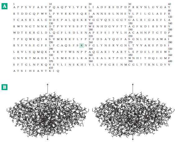
Plasma proteins have many functions. One of them, fullfilled principally by serum albumin, is to impart enough osmotic pressure to plasma to match that of the cytoplasm of cells. The heart-shaped human serum albumin consists of a single 65 kDa chain of 585 amino acid residues coiled into 28 helices. Three homologous repeat units or domains each contain six disulfide bridges, suggesting that gene duplication occurred twice during the evolution of serum albumins. The relatively low molecular mass and high density of negative charges on the surface make serum albumin well adapted for the role of maintaining osmotic pressure. However, serum albumin is not essential to life.


Over 50 mutant forms have been found and at least 30 persons have been found with no serum albumin in their blood. These analbuminemic individuals are healthy and have increased concentrations of other plasma proteins.
A second major function of plasma proteins is transport. Serum albumin binds to and carries many sparingly soluble metabolic products, including fatty acids, tryptophan, cysteine, steroids, thyroid hormones, Ca2+, Cu2+, Zn2+, other metal ions, bilirubin, and various drugs. There are also many more specialized transporter proteins. Transferrin carries iron and ceruloplasmin (an α2 globulin) transports copper. Transcortin carries corticosteroids and progesterone, while another protein carries sex hormones. Retinol-binding protein carries vitamin A and cobalamin-binding proteins vitamin B12. Hemopexin carries heme to the liver, where the iron can be recovered.j Haptoglobin binds hemoglobin released from broken red cells and also assists in the recycling of the iron in the heme.k Lipoproteins carry phospholipids, neutral lipids, and cholesterol esters. Most of the mass of these substances is lipid.
Immunoglobulins, α1-trypsin inhibitor and 2-macroglobulin, ten or more blood clotting factors; and proteins of the complement system all have protective functions that are discussed elsewhere in this book. Hormones, many of them proteins, are present in the blood as they are carried to their target tissues. Many serum proteins have unknown or poorly understood functions. Among these are the acute phase proteins, whose concentrations rise in response to inflammation or other injury.

пятница, 8 октября 2010 г.

Polypeptides

The chain formed by polymerization of amino acid molecules provides the primary structure of a protein. Together with any covalent crosslinkages and other modifications, this may also be called the covalent structure of the protein. Each monomer unit in the chain is known as an amino acid residue. This term acknowledges the fact that each amino acid has lost one molecule of H2O during polymerization. To be more precise, the number of water molecules lost is one less than the number of residues. Peptides are named according to the amino acid residues present and beginning with the one bearing the terminal amino group. Thus, L-alanyl-L-valyl-L-methionine has the following structure:


Like amino acids, this tripeptide is a dipolar ion. The same structure can be abbreviated Ala-Val-Met or, using one-letter abbreviations, AVM. It is customary in describing amino acid sequences to place the aminoterminal (N-terminal) residue at the left end and the carboxyl-terminal (C-terminal) residue at the right end. Residues are numbered sequentially with the N-terminal residue as 1.
The sequence of amino acid units in a protein is always specified by a gene. The sequence determines how the polypeptide chain folds and how the folded protein functions. For this reason much effort has gone into “sequencing,” the determination of the precise order of amino acid residues in a protein. Sequences of several hundreds of thousands of proteins and smaller peptides have been established and the number doubles each year.

(A) The complete amino acid sequence of the cytoplasmic enzyme aspartate aminotransferase from pig heart. The peptide has the composition Lys19, His8, Arg26, (Asp + Asn)42, Ser26, Thr26, (Glu + Gln)41, Pro24, Gly28, Ala32, Cys5, Val29, Met6, Ile19, Leu38, Tyr12, Phe23, Trp9. The molecular mass is 46.344 kDa and the complete enzyme is a 93.147-kDa dimer containing two molecules of the bound coenzyme pyridoxal phosphate attached to lysine-258 (enclosed in box).85,86 (B) A stereoscopic view of a complete enzyme molecule which contains two identical subunits with the foregoing sequence. Coordinates from Arthur Arnone. In this “wire model” all the positions of all of the nearly 7000 atoms that are heavier than hydrogen are shown. The > 8000 hydrogen atoms have been omitted. The view is into the active site of the subunit on the right. The pyridoxal phosphate and the lysine residue to which it is attached are shown with heavy lines. The active site of the subunit to the left opens to the back side as viewed here. The drawing may be observed best with a magnifying viewer available from Abrams Instrument Corp., Lansing, Michigan or Luminos Photo Corp., Yonkers, New York. However, with a little practice, it is possible to obtain a stereoscopic view unaided. Hold the book with good illumination about 20–30 cm from your eyes. Allow your eyes to relax as if viewing a distant object. Of the four images that are visible, the two in the center can be fused to form the stereoscopic picture. Drawings by program MolScript (Kraulis, 1991).


Most of these have been deduced from the sequences of nucleotides in DNA. Sequences of some small peptide hormones and antibiotics. The molecular mass of a protein can be estimated from the chain length by assuming that each residue adds 100–115 Da.
The amino acid composition varies greatly among proteins. A typical globular protein contains all or most of the 20 amino acids. The majority are often present in roughly similar amounts but His, Cys, Met, Tyr, and Trp tend to be less abundant than the others. Specialized proteins sometimes have unusual amino acid compositions. For example, collagen of connective tissue contains 33 mole% glycine and 21% of proline + hydroxyproline residues; the major proteins of saliva contain 22% of glutamate + glutamine and 20–45% proline. Cell walls of plants contain both high proline and high glycine polypeptides. One from petunias is 67% glycine. Silk fibroin contains 45% glycine and 29% alanine. A DNA repair protein of yeast has 13 consecutive aspartate residues. The tough eggshell (chorion) of the domesticated silkmoth Bombyx mori contains proteins with ∼30% cysteine. Many proteins consist, in part, of repeated short sequences. For example, the malaria-causing Plasmodium falciparum in its sporozoite stage is coated with a protein that contains 37 repeats of the sequence NANP interspersed with 4 repeats of NDVP. These two sequences have been indicated with single-letter abbreviations for the amino acids.
With a large number of protein and DNA sequences available, it has become worthwhile to compare sequences of the same protein in different species or of different proteins within the same or different species. Computer programs make it possible to recognize similarities and homologies between sequences even when deletions and insertions have occurred. The term homology has the precise biological definition “having a common evolutionary origin,” but it is often used to describe any close similarity in sequence. Among a pair of homologous proteins, a change at a given point in a sequence may be either conservative, meaning that a residue of similar character (large, small, positively charged, nonpolar, etc.) has been substituted, or it may be nonconservative.

понедельник, 4 октября 2010 г.

The Peptide Unit

The very ability of a protein to exist as a complex three-dimensional structure depends upon the properties of the amide linkages between the amino acid units. Many of these properties follow from the fact that an amide can be viewed as a resonance hybrid of the following structures. Because of the partial doublebond character, the C–N bond is shorter than that of a normal single bond and the C=O bond is lengthened.


The observed lengths in nanometers determined by X-ray diffraction measurements are given. The partial double-bond character of the C–N bond has important consequences. The peptide unit is nearly planar as is indicated by the dashed parallelogram.
However, the bonds around the nitrogen retain some pyramidal. Even more important is the fact that there is flexibility. As a result, the torsion angle ω may vary over a range of ± 15° or even more from that in the planar state. The resonance stabilization of the amide linkage is thought to be about 85 kJ/mol. Rotation around the C–N bond through 90° would be expected to require about this much energy. This fact immediately suggests a way in which proteins may sometimes be able to store energy—by having one or more peptide units twisted out of complete planarity.
Dimensions of the peptide linkage. Interatomic distances in nm, including the hydrogen bond length to an adjacent peptide linkage, are indicated. The atoms enclosed by the dotted lines all lie approximately in a plane. However, as indicated in the lower drawing, the nitrogen atom tends to retain some pyramidal character.


An important effect of the resonance of the amide linkage is that the oxygen atom acquires some negative charge and the NH group some positive charge. Some of the positive charge is usually depicted as residing on the nitrogen, but some is found on the hydrogen atom. The latter can be pictured as arising from a contribution of a fourth resonance form that contains no bond to hydrogen.


Nevertheless, this picture is inadequate. Various evidence indicates that the nitrogen actually carries a net negative charge.
The positive and negative ends of the dipoles in the amide group tend to associate to form strong hydrogen bonds. These hydrogen bonds together with the connecting amide linkages can form chains that may run for considerable distances through proteins. The tendency for cooperativity in hydrogen bond formation may impart unusual stability to these chains. As with individual amide linkages, these chains of hydrogen-bonded amides can also be thought of as resonance hybrids:


The two structures pictured are extreme forms, the true structure being something in between. In the lower form, rotation about the C–N bond would be permitted but then the charge separation present in the upper structure would no longer exist. Thus, the hydrogen bonds would be weakened. We can conclude that if an amide linkage in such a chain becomes twisted, the hydrogen bonds that it forms will be weakened. If there is cooperativity, the hydrogen bonds will all be strongest when there is good planarity in all of the amides in the chain.
Amides have very weak basic properties and protonation is possible either on the oxygen (A) or on the nitrogen (B).


The pKa values for such protonation are usually less than zero, but it is possible that a correctly placed acidic group in a protein could protonate either oxygen or nitrogen transiently during the action of a protein. Protonation on oxygen would strengthen hydrogen bonds from the nitrogen whereas protonation on nitrogen would weaken hydrogen bonds to oxygen and might permit rotation. The amide group has a permanent dipole moment of 3.63 debyes oriented as follows:


Here the arrow points toward the positive end of the dipole.