Fixed positive and negative charges attract each other strongly. Consider a carboxylate ion in contact with–NH3 + or with an ion of calcium:
From the van der Waals radii of Table 2-1 and the ionic crystal radius of Ca2+ of 0.10 nm, we can estimate an approximate distance between the centers of positive and negative charge of 0.25 nm in both cases. It is of interest to apply Coulomb’s law to compute the force F between two charged particles which are almost in contact. Let us choose a distance of 0.40 nm.
In this equation r is the distance in meters, q and q’ are the charges in coulombs (one electronic charge = 1.6021 x 10⁻¹⁹ coulombs), ε is the dielectric constant, and F is the force in newtons (N). The force per mole is NF where N is Avogadro’s number.
An uncertainty in this kind of calculation is in the dielectric constant ε, which is 1.0 for a vacuum, about 2 for hydrocarbons, and 78.5 for water at 25°C. If ε is taken as 2, the force for r = 0.40 nm is 4.3 x 10¹⁴ N/mol. The force would be twice as great for the Ca2+ – COO– case. To move two single charges further apart by just 0.01 nm would require 4.3 kJ/mol, a substantial amount of energy. However, if the dielectric constant were that of water, this would be reduced almost 40-fold and the electrostatic force would not be highly significant in binding. It is extremely difficult to assign a dielectric constant for use in the interior of proteins. For charges spaced far apart within proteins the effective dielectric constant is usually as high as 30–60. For closely spaced charges in hydrophobic niches it may be as low as 2–4.
A calculation that is often made is the work required to remove completely two charges from a given distance apart (e.g., 0.40 nm) to an infinite distance.
If ε = 2, this amounts to 174 kJ/mol for single charges at a distance of 0.40 nm; 69 kJ/mol at 1 nm; and only 6.9 kJ/mol at 10 nm, the distance across a cell membrane. We see that very large forces exist between closely spaced charges.
Electrostatic forces are of great significance in interactions between molecules and in the induction of changes in conformations of molecules. For example, attraction between –COO– and –NH3 + groups occurs in interactions between proteins. Calcium ions often interact with carboxylate groups, the doubly charged Ca2+ bridging between two carboxylate or other polar groups. This occurs in carbohydrates such as agarose, converting solutions of these molecules into rigid gels. Individual macromolecules as well as cell surfaces usually carry a net negative charge at neutral pH. This causes the surfaces to repel each other. However, at a certain distance of separation the van der Waals attractive forces will balance the electrostatic repulsion. Protruding hydrophobic groups may then interact and may“tether” bacteria or other particles at a fixed distance, often ~5 nm, from a cell surface.
среда, 25 августа 2010 г.
понедельник, 23 августа 2010 г.
Van der Waals Forces.
All atoms have a weak tendency to stick together,
and because of this even helium liquifies at a low
enough temperature. This is a result of the van der
Waals or “London dispersion forces” which act strongly only at a very short distance. These forces arise from electrostatic attraction between the positively charged nucleus of one atom and the negatively charged electrons of the other. Because nuclei are screened by the electron clouds surrounding them, the force is weak. The energy (enthalpy) of binding of one methylene (–CH2 –) unit into a monomolecular layer of a fatty acid is about –∆H° = 1.7 kJ/mol. 22 Although this is a small quantity, when summed over the 16 or more carbon atoms of a typical fatty acid the binding energy is substantial. When a methylene group is completely surrounded in a crystalline hydrocarbon, its van der Waals energy, as estimated from the heat of sublimation, is 8.4 kJ/mol; that of H2O in liquid water at the melting point of ice is 15 kJ/mol.
While van der Waals forces between individual atoms act over very short distances, they can be felt at surprisingly great distances when exerted by large molecules or molecular aggregates. Forces between very smooth surfaces have been measured experimentally at distances as great as 10 nm and even to 300 nm. However, these “long-range van der Waals forces” probably depend upon layers of oriented water molecules on the plates.
суббота, 21 августа 2010 г.
Forces between Molecules and between Chemical Groups.
The structure of living cells depends very much on the covalent bonds within individual molecules and on covalent crosslinks that sometimes form between molecules. However, weaker forces acting between molecules and between different parts of the same molecule are responsible for many of the most important properties of biochemical substances. These
are described as van der Waals forces, electrostatic forces, hydrogen bonds, and hydrophobic interactions. In the discussion that follows the thermodynamic quantities ∆H, ∆S, and ∆G will be used.
четверг, 19 августа 2010 г.
Tautomerism and Resonance.
Many simple organic compounds exist as mixtures of two or more rapidly interconvertible isomers or tautomeric forms. Tautomers can sometimes be separated one from the other at low temperatures where the rate of interconversion is low. The classic example is the oxo-enol (or keto-enol) equilibrium.
Although usually less stable than the oxo (keto) form, the enol is present in a small amount. It is formed readily from the oxo tautomer by virtue of the fact that hydrogen atoms attached to carbon atoms that are immediately adjacent to carbonyl (C=O) groups are remarkably acidic. Easy dissociation of a proton is a prerequisite for tautomerism. Since most hydrogen atoms bound to carbon atoms do not dissociate readily, tautomerism is unusual unless a carbonyl or other “activating group” is resent.
Since protons bound to oxygen and nitrogen atoms usually do dissociate readily, tautomerism also exists in amides and in ring systems containing O and N.
The tautomerism in is the counterpart of that in the oxo-enol transformation. However, the equilibrium constant for aqueous conditions favors form A very strongly. 2-Pyridone is tautomerized to 2-hydroxypyridine to a greater extent. Pyrimidines and purines can form a variety of tautomers. The existence of form D of is the basis for referring to uracil as dihydroxypyrimidine. However, the di-oxo tautomer A redominates. Pyridoxine (vitamin B6) exists in water largely as the dipolar ionic tautomer B but in methanol as the uncharged tautomer A. In a pair of tautomers, a hydrogen atom always moves from one position to another and the lengths and bond haracter of these bonds also change.
The equilibrium constant for a tautomeric interconversion is simply the ratio of the mole fractions of the two forms; for example, the ratio of enol to oxo forms of acetone12 in water at 25°C is 6.0 x 10⁻⁹, while that for isobutyraldehyde is 1.3 x 10⁻⁴. The ratio of 2-hydroxypyridine to 2-pyridone is about 10–3 in water but increases to 0.6 in a hydrocarbon solvent and to 2.5 in the vapor phase. The ratio of dipolar ion to uncharged pyridoxine is ∼4 at 25°C in water. The ratios of tautomers B, C, and D to the tautomer A of uracil are small, but it is ifficult to measure them quantitatively. These tautomeric ratios are defined for given overall states of rotonation. The constants are independent of pH but will change if the overall state f protonation of the molecule is changed. They may also be altered by changes in temperature or solvent or by binding to a protein or other molecule.
It is important to distinguish tautomerism from resonance, a term used to indicate that the properties of a given molecule cannot be represented by a single valence structure but can be represented as a hybrid of two or more structures in which all the nuclei remain in the same places. Only bonding electrons move to convert one resonance form into another. Examples are the enolate anion, which can be thought of as a hybrid of structures A and B, and the amide linkage, which can be represented by a similar pair of resonance forms.
A double-headed arrow is often used to indicate that two structures drawn are resonance structures rather than tautomers or other separable isomers.
Although they are distinctly different, tautomerism and resonance are related. Thus, the acidity of carbon-bound hydrogens in ketones, which allows formation of enol tautomers, results from the fact that the enolate anion produced by dissociation of one of these hydrogens is stabilized by resonance. Similarly, tautomerism in the imidazole group of the amino acid histidine is related to resonance in the imidazolium cation. Because of this resonance, if a proton approaches structure A of and becomes attached to the lefthand nitrogen atom (Nδ), the positive charge in the resulting intermediate is distributed over both nitrogen atoms. This makes the proton on Nε acidic, permitting it to dissociate to tautomer B.
Since Nσ has sometimes also been called N3, it is best not to use the numerical designations for the nitrogen atoms. The tautomeric ratio of B to A for histidine in
water has been estimated, using 15N- and 13C-NMR, as 5.0 when the α-amino group is protonated and as 2.5 when at high pH it is unprotonated. This tautomerism of the imidazole group is probably important to the function of many enzymes and other proteins; for example, if Nε of structure A is embedded in a protein, a proton approaching from the outside can induce the tautomerism shown with the release of a proton in the interior of the protein, perhaps at the active site of an enzyme. The form protonated on Nδ, which is the minor form in solution, predominates in some positions within proteins.
вторник, 17 августа 2010 г.
Conformations: The Shapes That Molecules Can Assume.
As important to biochemists as configurations, the stable arrangements of bonded atoms, are conformations, the various orientations of groups that are caused by rotation about single bonds. In many molecules such rotation occurs rapidly and freely at ordinary temperatures. We can think of a –CH3 group as a kind of erratic windmill, turning in one direction, then another. However, even the simplest molecules have preferredconformations, and in more complex structures rotation is usually very restricted.
Consider a molecule in which groups A and B are joined by two CH2 (methylene) groups. If A and B are pulled as far apart as possible, the molecule is in its fully extended anti or staggered conformation:
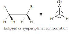
Groups A and B are said to be antiperiplanar (ap) in this conformation. Not only are A and B as far apart as possible but also all of the hydrogen atoms are at their maximum distances one from the other. This can be seen by viewing the molecule down the axis joining the carbon atoms (Newman projection). Rotation of the second carbon atom 180° around the single bond yields the eclipsed conformation in which groups A and B are synperiplanar.
If A and B are large bulky groups they will bump together, attainment of the eclipsed conformation will be almost impossible, and rotation will be severely restricted. Even if A and B are hydrogen atoms (ethane), there will be a rotational barrier in the eclipsed conformation which amounts to ∼12 kJ (3 kcal) per mole because of the crowding of the hydrogen atoms as they pass each other. This can be appreciated readily by examination of space-filling molecular models.
If groups A and B are methyl groups (butane), the steric hindrance between A and B leads to a rotational barrier of ∼25 kJ ( 6 kcal) per mole. The consequence of this simple fact is that in fatty acids and related substances and in polyethylene the chains of CH2 groups tend to assume fully extended zigzag conformations.
In addition to this extended conformation there are two gauche (skewed or synclinal) conformations which are only slightly less stable than the staggered conformation and in which A and B interfere only if they are very bulky. In one of the twogauche conformations B lies to the right of A and in the other to the left of A when viewed down the axis.
These two conformations are related to right-handed and left-handed screws, respectively. The threads on an ordinary right-handed household screw, when viewed down the axis from either end, move backward from left to right in the same fashion as do the groups A and B in the illustration. The angle φ is the torsion angle and is positive for right-handed conformations. Gauche conformations are important in many biological molecules; for example, the sugar alcohol ribitol stacks in crystals in a “sickle” conformation, in which the chain starts out (at the left) in the zigzag arrangement but shifts to a gauche conformation around the fourth carbon atom, thereby minimizing steric interference between the OH groups on the second and fourth carbons.
The complete series of possible conformations is shown
in Fig 1.
In the chain of methylene units, the hydrogen atoms on alternate carbon atoms of the fully extended chain barely touch but larger atoms cannot be accommodated. Thus, when fluorine atoms of van der Waals radius 0.135 nm replace the hydrogen atoms of radius 0.12 nm, a fully extended chain is no longer possible. For this reason the torsion angle in polyfluoroethylene is changed from the 180° of polyethylene to 166°, enough to relieve the congestion but not enough to cause severe eclipsing of the fluorines on djacent carbons. The resulting helical structure is reminiscent of those occurring in proteins and other biopolymers. We see that helix formation can be a natural result of steric hindrance between groups of atoms.
Consider a molecule in which groups A and B are joined by two CH2 (methylene) groups. If A and B are pulled as far apart as possible, the molecule is in its fully extended anti or staggered conformation:
Groups A and B are said to be antiperiplanar (ap) in this conformation. Not only are A and B as far apart as possible but also all of the hydrogen atoms are at their maximum distances one from the other. This can be seen by viewing the molecule down the axis joining the carbon atoms (Newman projection). Rotation of the second carbon atom 180° around the single bond yields the eclipsed conformation in which groups A and B are synperiplanar.
If A and B are large bulky groups they will bump together, attainment of the eclipsed conformation will be almost impossible, and rotation will be severely restricted. Even if A and B are hydrogen atoms (ethane), there will be a rotational barrier in the eclipsed conformation which amounts to ∼12 kJ (3 kcal) per mole because of the crowding of the hydrogen atoms as they pass each other. This can be appreciated readily by examination of space-filling molecular models.
If groups A and B are methyl groups (butane), the steric hindrance between A and B leads to a rotational barrier of ∼25 kJ ( 6 kcal) per mole. The consequence of this simple fact is that in fatty acids and related substances and in polyethylene the chains of CH2 groups tend to assume fully extended zigzag conformations.
In addition to this extended conformation there are two gauche (skewed or synclinal) conformations which are only slightly less stable than the staggered conformation and in which A and B interfere only if they are very bulky. In one of the twogauche conformations B lies to the right of A and in the other to the left of A when viewed down the axis.
These two conformations are related to right-handed and left-handed screws, respectively. The threads on an ordinary right-handed household screw, when viewed down the axis from either end, move backward from left to right in the same fashion as do the groups A and B in the illustration. The angle φ is the torsion angle and is positive for right-handed conformations. Gauche conformations are important in many biological molecules; for example, the sugar alcohol ribitol stacks in crystals in a “sickle” conformation, in which the chain starts out (at the left) in the zigzag arrangement but shifts to a gauche conformation around the fourth carbon atom, thereby minimizing steric interference between the OH groups on the second and fourth carbons.
The complete series of possible conformations is shown
in Fig 1.
Figure 1. Description of conformations about a single bond in the terminology of Klyne and Prelog10,11 using the Newman projection. Group A is on the front atom at the top: the conformation is given for each possible position of group B on the other atom.
In the chain of methylene units, the hydrogen atoms on alternate carbon atoms of the fully extended chain barely touch but larger atoms cannot be accommodated. Thus, when fluorine atoms of van der Waals radius 0.135 nm replace the hydrogen atoms of radius 0.12 nm, a fully extended chain is no longer possible. For this reason the torsion angle in polyfluoroethylene is changed from the 180° of polyethylene to 166°, enough to relieve the congestion but not enough to cause severe eclipsing of the fluorines on djacent carbons. The resulting helical structure is reminiscent of those occurring in proteins and other biopolymers. We see that helix formation can be a natural result of steric hindrance between groups of atoms.
воскресенье, 15 августа 2010 г.
Geometrical isomers.
The RS system also gives an unambiguous designation of geometrical isomers containing a double bond. At each end of the bond, select the group of highest priority. If these two groups lie on the same side of the double bond the configuration is Z (from the German zusammen,“together”); if on opposite sides E (entgegen,“opposite”).
Configurations of amide or ester linkages may also be specified in this manner. This is possible because the C–N bond of an amide has partial double-bond character, as to a lesser extent does the C–O bond to the bridge oxygen in an ester. In this case, assign the lowest priority to the unshared electron pair on the ester bridge oxygen.
An amide of the Z configuration is ordinarily referred to as trans in protein chemistry because the main chain atoms are trans.
пятница, 13 августа 2010 г.
Diastereoisomers.
Whereas compounds with one chiral center exist as an enantiomorphic pair, molecules with two or more chiral centers also exist as diastereoisomers (diastereomers). These are pairs of isomers with an opposite configuration at one or more of the chiral centers, but which are not complete mirror images of each other. An example is L-threonine which has the 2S, 3R configuration. The diastereoisomer with the 2S, 3S configuration is known as L-allo-threonine. L-isoleucine, whose side chain is –CH(CH3) CH2CH3, has the 2S, 3R configuration. It can be called 2(S)- amino-3(R)-methyl-valeric acid but the simpler name L-isoleucine implies the correct configuration at both chiral centers.
Sometimes the subscript s or g is added to a D or L prefix to indicate whether the chirality of a compound is being related to that of serine, the traditional configurational standard for amino acids, or to that of glyceraldehyde. In the latter case the sugar convention (Chapter 4) is followed. In this convention the configurations of the chiral centers furthest from C1 are compared. Ordinary threonine is Ls- or Dg-threonine. The configuration of dextrorotatory (+)-tartaric acid can be described as 2R, 3R, or as Ds, or as Lg.
Biochemical reactions are usually stereospecific and a given enzyme will catalyze reactions of molecules of only a single configuration. A related fact is that proteins ordinarily consist entirely of amino acids of the L series.
среда, 11 августа 2010 г.
The RS notation for configuration.
This notation, devised by Cahn, Ingold, and Prelog, provides an unambiguous way of specifying configuration at any chiral center. It is especially useful for classes of compounds for which no well-established DL system is available. The groups or atoms surrounding the central carbon atom, or other central atom, are ranked according to a prioritysequence. The priority of a group is determined by a number of sequence rules, the first of which is (1) Higher atomic number precedes lower. In the following illustration, the priorities of the groups in D-alanine are indicated by the letters a > b > c > d. The highest priority (a) is assigned to the NH2 groups which contain nitrogen bonded to the central atom. To establish the configuration, the observer views the molecule down the axis connecting the central atom to the group having the lowest priority, i.e., to group d. Viewed in this way, the sequence of groups a, b, and c can either be that of a right-handed turn (clockwise) as shown in the drawing or that of a left-handed turn (counterclockwise).
To establish the priority sequence of groups first look at the atoms that are bonded directly to the central atom, arranging them in order of decreasing atomic number. Then if necessary, move outward to the next set of atoms, again comparing atomic numbers. In the case of alanine, groups b and c must be ordered in this way because they both contain carbon directly bonded to the central atom. When double bonds are present at one of the atoms being examined, e.g., the carboxyl group in alanine, imagine that phantom atoms that replicate the real ones are present at the ends of the bonds:
These phantom atoms fill out the valences of the atoms involved in the multiple bonds and are considered to have zero atomic number and zero mass. They are not considered in establishing priorities.
If the first rule and the expansion of multiple bonds are not sufficient to establish the priority, use these additional rules: (2) Higher atomic mass precedes lower. (3) When a double bond is present Z precedes E (see Geometrical isomers). For ring systems a cis arrangement of the highest priority substituents precedes trans. (4) When a pair of chiral centers is present R,R or S,S precedes R,S or S,R. (5) An R chiral center precedes S. For further details see Eliel et al. and Bentley. The following groups are ordered in terms of decreasing priority6: SH > OR > OH > NH–COCH3 > NH2 > COOR > COOH > CHO > CH2OH > C6H5 > CH3 > 3H > 2H > H.
Although the RS system is unambiguous, closely related compounds that belong to the same configurational family in the DL system may have opposite configurations in the RS system. Thus, L-cysteine (side chain–CH2SH) has the R configuration. This is one of the reasons that the DL system is still used for amino acids and sugars.
The view down the axis and toward the group of lowest priority (d), which lies behind the page. The right-handed turn indicates the configuration R (rectus = right); the opposite configuration is S (sinister = left).
To establish the priority sequence of groups first look at the atoms that are bonded directly to the central atom, arranging them in order of decreasing atomic number. Then if necessary, move outward to the next set of atoms, again comparing atomic numbers. In the case of alanine, groups b and c must be ordered in this way because they both contain carbon directly bonded to the central atom. When double bonds are present at one of the atoms being examined, e.g., the carboxyl group in alanine, imagine that phantom atoms that replicate the real ones are present at the ends of the bonds:
These phantom atoms fill out the valences of the atoms involved in the multiple bonds and are considered to have zero atomic number and zero mass. They are not considered in establishing priorities.
If the first rule and the expansion of multiple bonds are not sufficient to establish the priority, use these additional rules: (2) Higher atomic mass precedes lower. (3) When a double bond is present Z precedes E (see Geometrical isomers). For ring systems a cis arrangement of the highest priority substituents precedes trans. (4) When a pair of chiral centers is present R,R or S,S precedes R,S or S,R. (5) An R chiral center precedes S. For further details see Eliel et al. and Bentley. The following groups are ordered in terms of decreasing priority6: SH > OR > OH > NH–COCH3 > NH2 > COOR > COOH > CHO > CH2OH > C6H5 > CH3 > 3H > 2H > H.
Although the RS system is unambiguous, closely related compounds that belong to the same configurational family in the DL system may have opposite configurations in the RS system. Thus, L-cysteine (side chain–CH2SH) has the R configuration. This is one of the reasons that the DL system is still used for amino acids and sugars.
понедельник, 9 августа 2010 г.
The D- and L- families of amino acids.
The amino acids of which proteins are composed are related to L-alanine but have various side chains (R groups) in place of the methyl group of alanine. In the preceding section the structure of L-alanine was given in four different ways. To recognize them all as the same structure, we can turn them in space to an orientation in which the carboxyl group is at the top, the side chain (–CH3) is down, and both project behind the paper. The amino group and hydrogen atom will then project upward from the paper at the sides as shown below. According to a convention introduced at the beginning of this century by Emil Fischer, an amino acid is L if, when oriented in this manner, the amino group lies to the left and D if it lies to the right.
Fischer further proposed that the amino acid in this orientation could be projected onto the paper and drawn with ordinary lines for all the bonds. This gives the previously shown Fischer projection formula of L-alanine.
Although the D and L system of designating configuration is old it is still widely used. Remember that D and L refer to the absolute configurations about a selected reference atom in the molecule; for an amino acid this is the number 2 or α-carbon. A quantity that is related to the asymmetry of molecules is the experimentally measurable optical rotation. The sign of the optical rotation (+ or –) is sometimes given together with the name of a compound, e.g., D(+)-glucose. The older designations d (dextro) and l (levo) indicated + and–, respectively. However, compounds with the D configuration may have either + or – optical rotation.
In older literature optical isomerism of the type represented by D and L pairs was usually discussed in terms of “asymmetric carbon atoms” or “asymmetric centers.” Now the terms chiral (pronounced ki-ral) molecules, chiral centers, and chirality (Greek: “handedness”) are preferred.
суббота, 7 августа 2010 г.
Asymmetry: Right-Handed and Left- Handed Molecules.
The left hand looks much like the right hand, but they are different. One is the mirror image of the other. A practical difference is that your right hand will not fit into a left-handed glove. Despite our daily acquaintance with “handedness” it may seem difficult to explain in words how a right and a left hand differ. However, since most biochemical compounds are asymmetric, it is important to be able to visualize these molecules in three dimensions and to draw their structures on paper. One of the best ways of learning to do this is to study molecular models. You may learn the most by making your own models (see
Appendix).
Whenever four different groups are bonded to a central carbon atom, the molecule is asymmetric and the four groups can be arranged in two different configurations. Consider alanine, one of the alpha (α)-amino acids from which proteins are built.
It is called an α-amino acid because the amino group is attached to the α carbon atom. To indicate its three-dimensional structure on a flat piece of paper, the bonds that project out of the plane of the paper and up toward the reader are often drawn as elongated triangles, while bonds that lie behind the plane of the paper are shown as dashed lines. The isomer of alanine having the configuration about the α-carbon atom shown in the following structural formulas is called S-alanine or L-alanine. The isomer
which is a mirror image of S-alanine is R-alanine or D-alanine. Pairs of R and S compounds (see Section B for definitions) are known as enantiomorphic forms or enantiomers.
Notice that in the foregoing drawings, the carboxyl
group, abbreviated –COOH-, is shown as havinglost a proton to form the carboxylate ion –COO–. Likewise, the amino group (–NH2) has gained a proton to form the –NH3 + ion. The resulting dipolar ionic or zwitterionic structure is the one that actually exists for amino acids both in solution and in crystals.
Подписаться на:
Сообщения (Atom)